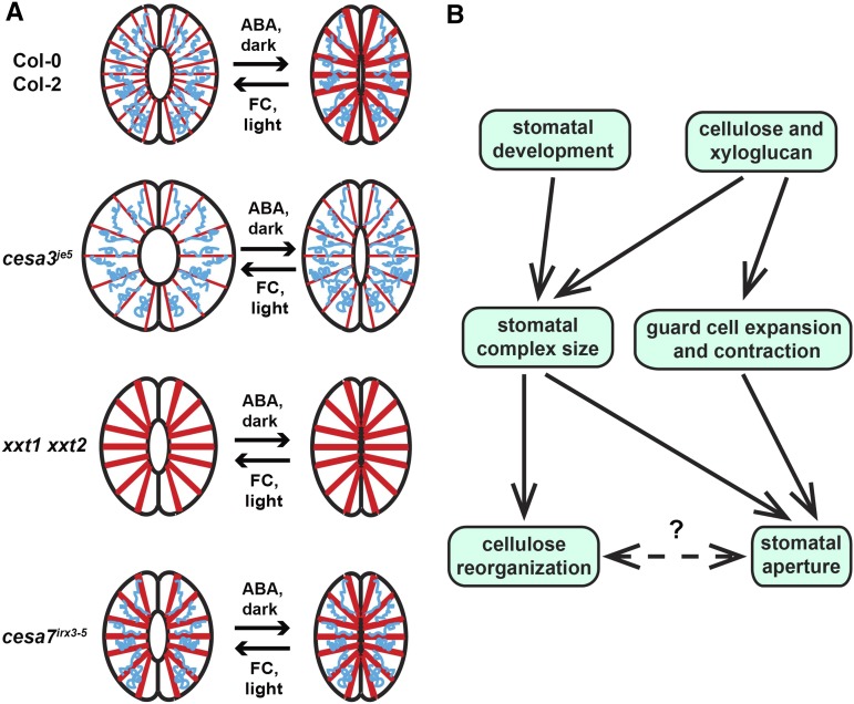
Figure 7.
Models for cellulose and xyloglucan in guard cells during stomatal movement. A, Schematics summarizing stomatal aperture changes and cellulose organization patterns in wild-type, cesa3je5, xxt1 xxt2, and cesa7irx3-5 stomata. Cellulose is represented as red spokes, and xyloglucan is demonstrated as blue coils that interact with cellulose microfibrils at limited regions. With sufficient amounts of cellulose and xyloglucan present in guard cells, cellulose reorganizes between a more homogenously distributed pattern in the open state and a more fibrillar, bundled pattern in the closed state. In the case of cellulose deficiency, stomatal aperture is larger because changes in guard cell length occur more readily. Cellulose reorganization is less evident, with more evenly spaced microfibrils in both open and closed states. In the absence of detectable xyloglucan, stomatal aperture is relatively smaller due to more restricted guard cell expansion and contraction, and cellulose is more bundled in both open and closed states. In cesa7irx3-5 mutants, the overall size of the stomatal complex is smaller (Liang et al., 2010), but cellulose is not deficient in guard cells, with fibrillar patterns in both states. B, Conceptual model illustrating a possible scenario of how cellulose and xyloglucan in guard cell walls function in the regulation of stomatal aperture and cellulose reorganization.

