Figure 3.
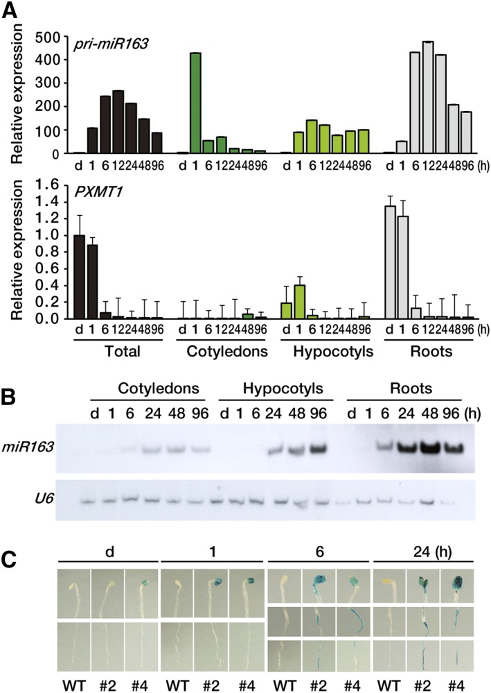
Tissue-specific expression pattern of miR163 and PXMT1. A, Expression levels of pri-miR163 and PXMT1 in various tissues during seedling de-etiolation. Seedlings were dissected into three parts (cotyledons, hypocotyls, and roots), and samples were collected indicated. Expression levels were determined by qRT-PCR. The values were normalized to that of the d sample of each panel. Error bars indicate sd (n = 3). B, Small RNA gel-blot analysis showing miR163 expression in cotyledons, hypocotyls, and roots. Total RNA was extracted from the samples described in A. U6 snRNA levels were used as a loading control. C, Histochemical localization of GUS activity in transgenic Arabidopsis seedling carrying the pri-miR163 promoter::GUS-GFP transgene. Four-day-old etiolated seedlings (d) were treated with W light as indicated (1, 6, and 24 h). WT, Wild type.

