Figure 2.
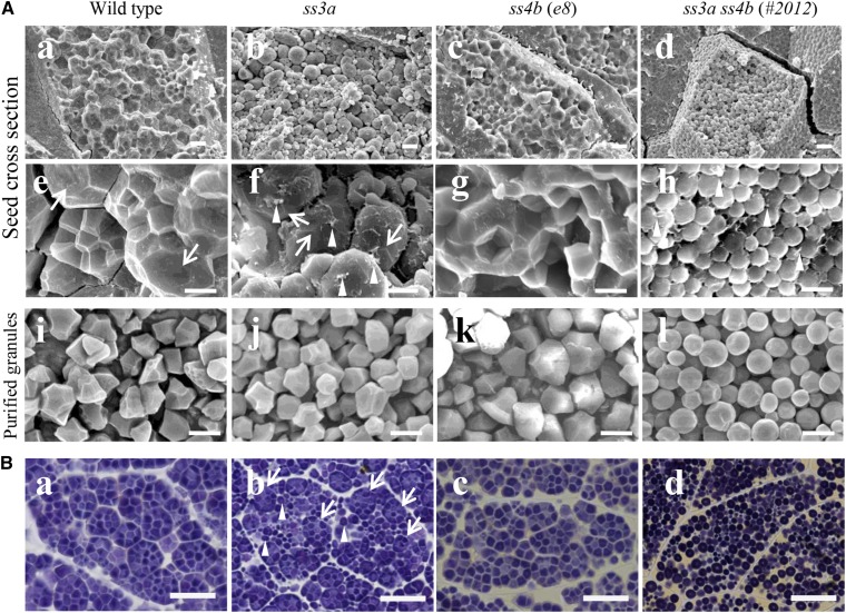
Starch granule morphology in mature rice seeds. A, Scanning electron micrographs of cross sections of mature seeds (central part of the endosperm; a–h) and purified starch granules (i–l). Arrows indicate the amyloplast surface, and arrowheads indicate putative shreds of the amyloplast envelope. Bars = 10 μm (a–d) and 5 μm (e–l). B, Light microscopy of iodine-stained thin cross sections of mature seeds (central part of the endosperm). Arrows indicate compound-type granules, and arrowheads indicate spherical granules. Bars = 20 μm.

