Figure 3.
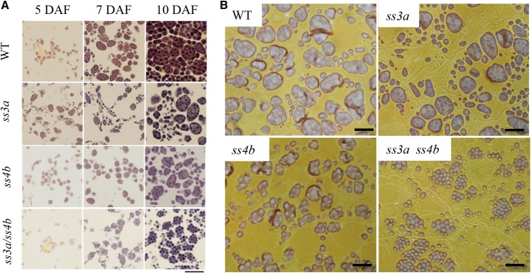
Starch granule morphology in developing endosperm. Light microscopy shows thin iodine-stained sections of the central part of the developing endosperm. Wild-type cv Nipponbare (WT), ss3a, ss4b (e8), and ss3a ss4b (#2012) were compared. A, Developing endosperm at 5, 7, and 10 DAF. Bar = 20 μm. B, Developing endosperm at 7 DAF. Bars = 10 μm.

