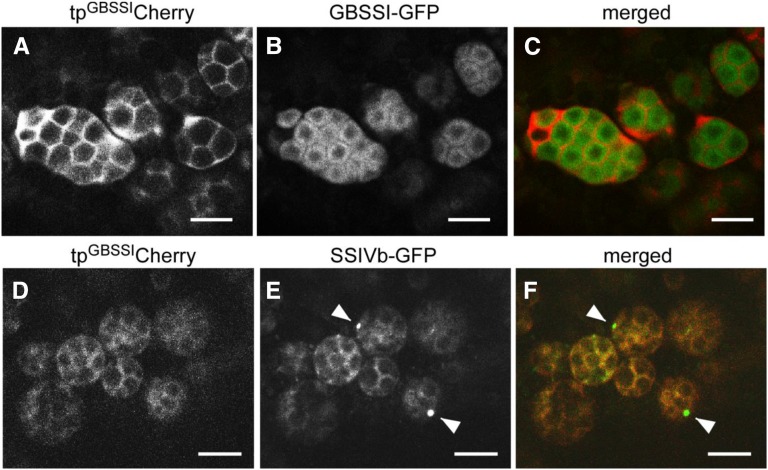
Figure 5.
Distinct localizations of GBSSI and SSIVb. Confocal fluorescence microscopy shows developing seeds of transformed wild-type (cv Yukihikari) plants (7 DAF) expressing tpGBSSICherry together with either GBSSI-GFP (A–C) or SSIVb-GFP (D–F). A to C, tpGBSSICherry (magenta channel) and GBSSI-GFP (green channel). Note that the two signals do not overlap, indicating that tpGBSSICherry is not efficiently imported into the stroma but is located primarily in the SLS and envelope. D to F, tpGBSSICherry (magenta channel) and SSIVb-GFP (green channel). Note that the SSIVb-GFP and tpGBSSICherry signals overlap extensively and that SSIVb-GFP is also detected as dots (arrowheads). Bars = 5 μm.

