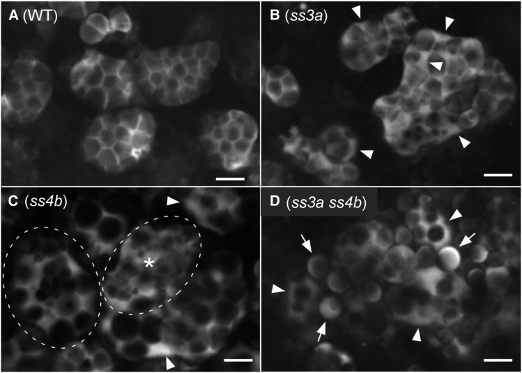
Figure 6.
Internal structures of amyloplasts visualized with tpGBSSIGFP. Developing seeds of the transformed wild-type (WT) cv Nipponbare (A), ss3a (B), ss4b (e14; C), and ss3a ss4b (#2013; D) were analyzed at 7 DAF by confocal microscopy. In B and C, arrowheads indicate enlarged IMS between the OEM and the IEM. In C, an amyloplast indicated by a dashed oval and asterisk does not contain a typical SLS lattice structure. Another amyloplast, indicated by a dashed oval, contains a cluster of small granules in the center and large granules at the periphery. Note that these distinct phenotypes in ss4b were observed with low frequency (less than 10%). Most amyloplast phenotypes were similar to that of the wild type. In D, arrowheads indicate amyloplasts containing multiple starch granules and arrows indicate granules that look like simple-type grains. Bars = 5 μm.

