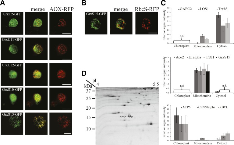
Figure 1.
Subcellular localization of Grx. A, Expression of Grx-GFP fusion constructs in Arabidopsis cell suspension culture. GFP signal (left), mitochondrial marker AOX-RFP (right), and the merged image (middle). Bar = 20 µm. B, Expression of GrxS15-GFP fusion construct in Arabidopsis cell suspension culture. GFP signal (left), plastid marker RbcS-RFP (right), and the merged image (middle). Bar = 20 µm. C, MRM analysis using specific peptides for GrxS15 and marker proteins for cytosol (top), mitochondria (middle), and chloroplast (bottom) in cell fractions from 2-week-old wild-type leaves. Top: GAPC2 (At1g13440), LOS1 (At1g56070), Trxh3 (At5g42980). Middle: Aco2 (At2g05710), E1α (At1g59900), PDH (At1g24180), GrxS15 (At3g15660). Bottom: ATP6 (AtCg00480), CPN60α (At2g28000), RBCL (AtCg00490). D, Mitochondrial protein of green tissue separated on a 2D Tricine gel with a 4–7 pI strip as the first dimension. Spots identified as GrxS15 are circled. The unmodified form is highlighted with a star.

