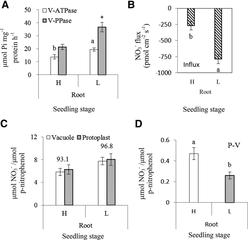
Figure 1.
B. napus with higher NUE showed lower VSC for NO3− in roots at the seedling stage. H refers to the high-NUE oilseed rape genotype Xiangyou15 and L refers to the low-NUE genotype 814. Specific activities of the tonoplast proton pumps are expressed as μmol Pi released mg−1 protein h−1. NO3− fluxes are expressed as pmol NO3− cm−2 S−1. Mature vacuoles were collected from the root tissues at seedling stage and a microelectrode was vibrated in the measuring solution between the two positions 1 μm and 11 μm from the vacuole surface (tonoplast) along an axis perpendicular to the tangent of the target vacuoles recording the stable reading data. The background was recorded by vibrating the electrode in measuring solution without vacuoles. Protoplasts and vacuoles isolated from roots of hydroponically grown plants were measured for NO3− content and NO3− accumulation normalized against the specific activity of the vacuole acid phosphatase (ACP) as described in “Materials and Methods”; and plotted as μmol NO3− per μmol p-nitrophenol, the end product of ACP assay. Proton pump activities in root tissues between H and L genotypes are shown at the seedling stage (A). Different letters at the top of the histogram bars denote significant differences of V-ATPase in root tissues between H and L genotypes (P < 0.05); an asterisk (*) at the top of the histogram bars indicates significant difference in V-PPase activity in root tissues between H and L genotypes (P < 0.05). Vertical bars on the figures indicate SD (n = 6). Mean rates of NO3− flux during 160 s within vacuoles of root tissues between H and L genotypes are shown at the seedling stage (B). Different letters at the top of the histogram bars denote significant differences of NO3− flux between H and L genotypes (P < 0.05). Vertical bars on the figures indicate SD (n = 6). Accumulation of NO3− inside the vacuole and in the protoplasts of root tissues is shown at the seedling stage (C). Values above the bars represent the percentage of vacuolar NO3− relative to the total NO3− in protoplasts. NO3− accumulation in the cytosol of root tissues is shown at seedling stage (D). P-V is the total NO3− in the cytosol and was calculated a tostal NO3− in protoplasts – total NO3− in vacuole. Different letters at the top of histogram bars denote significant differences of total NO3− in cytosol between H and L genotypes (P < 0.05). Vertical bars on the figures indicate SD (n = 6).

