Figure 3.
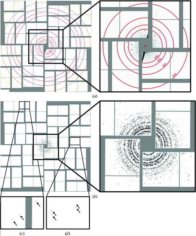
(a) Example SFX diffraction image of ΔPEX11 cells displaying Bragg-sampled reflections with intensities above the background level. (b) Composite XRPD patterns assembled from individual diffraction images show that most crystallites diffract to approximately 30 Å resolution, with several crystals displaying diffraction out to the detector edge (6 Å) and corners (5.6 Å) as indicated by arrows in insets (c) and (d).

