Fig. 3.
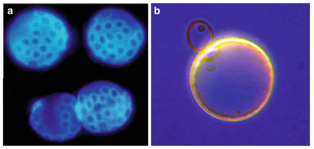
Distinguishing old yeast cells from young. (a) Old yeast cells that have been sorted using magnetic sorting and stained with calcofluor dye. Each blue stained ring is a “bud scar,” a deposit of chitin that remains on the surface of the cell wall at each site of division (photo credit James Claus). (b) Young and old yeast cells stained with bromophenol blue, showing the larger size and thicker cell wall of the old mother cell

