Figure 3.
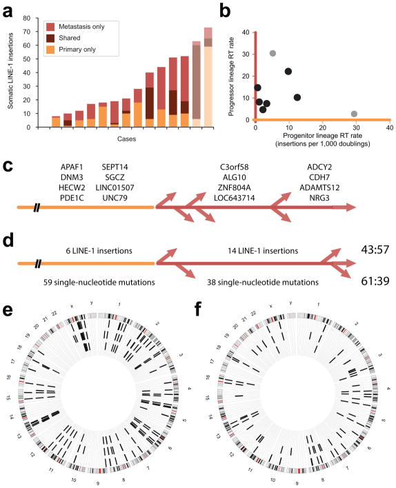
Retrotransposition (RT) events. (a) Somatic LINE-1 insertions. Stacked bars show insertions found in only the primary tumor (orange, bottom); those shared by the primary and metastatic samples (middle); and those only in the metastasis (red). Confirmed PDAC cases are shown in colors indicated by the legend; two other gastrointestinal adenocarcinomas are shown in lighter hues. (b) RT rate was calculated for each case during two phases of clonal evolution. The progenitor lineage rate (orange, x-axis) is proportional to the number of LINE-1 insertions acquired within a cellular lineage in the primary tumor antedating its seeding a metastatic site. These are shared by the primary and metastatic sites. The progressor lineage rate (red, y-axis) reflects insertions found only in the metastatic sample. PDAC cases are indicated with black dots; other adenocarcinomas are in grey. (c) Somatically acquired LINE-1 in PDAC. Twenty insertions were detected in the primary and metastatic samples for this case and not in normal DNA. Gene names are listed for 8 intronic insertions. These occurred in the progenitor lineage of the primary tumor (leftmost, orange); they were PCR amplified from all metastases. Eight insertions were found by TIP-seq at a metastatic site and not in the primary tumor; nearest gene names are given for intergenic insertions. Four were present in two additional metastases and occurred in a progressor clone of the primary tumor (middle, red). The remaining four were unique to the metastasis profiled by TIP-seq (rightmost) and occurred later in the primary tumor or at that metastatic site. (d) Somatically acquired LINE-1 and single-nucleotide mutations in PDAC. Numbers of events occurring in the progenitor (orange) and progressor (red) lineages are shown; ratios are on the right. (e) LINE-1 insertions in a gastric adenocarcinoma. Sixty insertions were detected in the primary tumor; most (54) were shared with the metastatic site. (f) LINE-1 insertions in a duodenal adenocarcinoma. Twenty-seven insertions were identified in the primary, and 42 in the metastasis. Eighteen were shared between the two sites.

