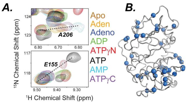
Figure 3. CONCISE analysis of the chemical shift changes with nucleotide.
A) [1H,15N]-TROSY-HSQC spectra showing the backbone amide chemical shift changes of PKA-C saturated with different nucleotides upon binding PKI5-24 (See also Figure S4–6) Residues following linear trajectories (blue spheres) plotted on the cartoon representation of the PKA-C crystal structure (PDB: 1ATP).

