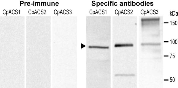
Figure 4.
Western blot analysis of the three CpACS proteins. Crude protein extracts were prepared from free C. parvum sporozoites by in vitro excystation. Primary antibodies were affinity-purified. Antibodies from pre-immune animals were also affinity-purified and used as control. For clarity and easy comparison, images from the three membrane strips were digitally scaled according to positions of protein standards. The region corresponding to the CpACS bands were indicated by an arrowhead.

