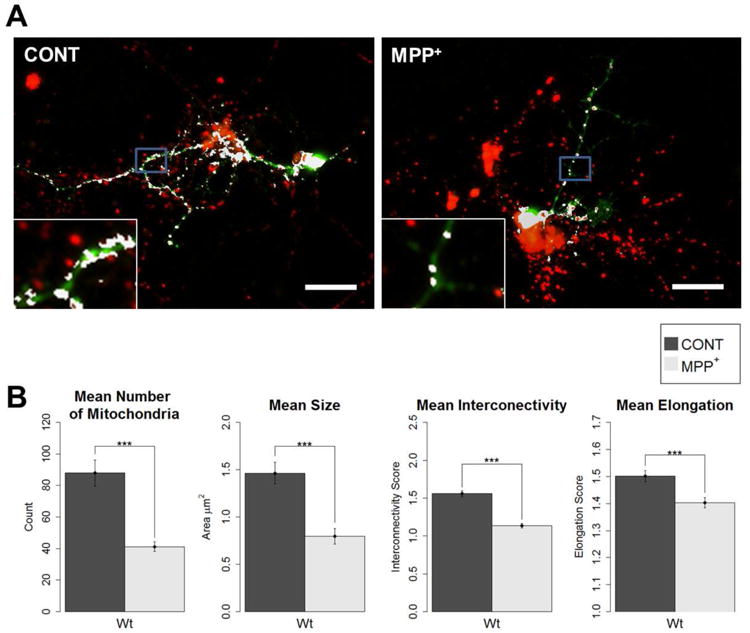
Figure 4. MPP+ treatment causes fragmented mitochondrial morphology.

Primary neuronal cell cultures were prepared from wild-type fly embryos. At 3 DIV, cultures were treated with 40 μM MPP+, or as control. At 7DIV, cells were stained with anti-TH and MitoTracker for the colocalization analysis of mitochondria in DA neurons. A) Example images of control and MPP+-treated dopaminergic (DA) neurons from immunocytochemistry colocalization method (anti-TH and MitoTracker). Mitochondria of dopaminergic neurons are shown in white (scale bar = 20 μm, insets are of areas outlined in blue). B) All four parameters showed a significant decrease from control when treated with MPP+. Primary neuronal cell cultures were prepared from wild-type fly embryos. At 3 DIV, cultures were treated with 40 μM MPP+, or as control. At 7DIV, cells were stained with MitoTracker and anti-TH for the colocalization analysis, and images were taken for analysis (Student's t-test; *** p < 0.001); number of DA neurons analyzed: Control = 29, MPP+ = 51).
