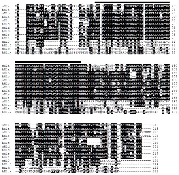
Fig. 1. Multiple alignment of mouse and human somatic H1 variants.
Genbank accession numbers of the protein sequences are listed in Table 1. The black line on top of amino acid sequences marks the globular domain. The conserved and the similar residues are highlighted in black and grey colors, respectively.

