Figure 2.
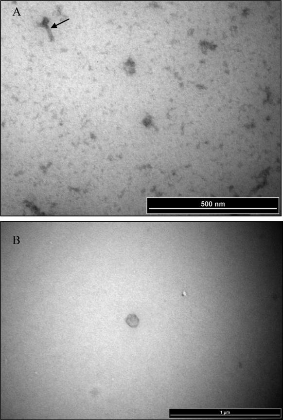
Representative electron micrograph images of phage particles isolated and purified from F.nucleatum strain 7_1. Panels A) ɸFunu1; and B) ɸFunu2.
Arrow in panel A indicates a putative phage tail structure. Note the absence of a tail structure for the representative virion head imaged in panel B.

