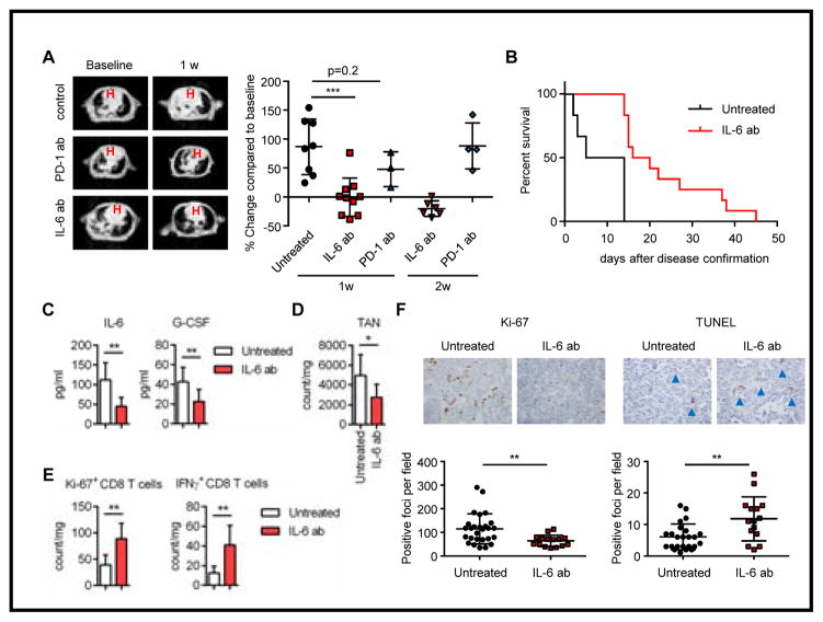
Figure 4. IL-6 neutralizing treatment showed clinical efficacy in Kras/Lkb1 mouse model.
A. Representative images of Magnetic resonance imaging (MRI) and quantification of MRI from KL mice treated with PD-1 or IL-6 blocking antibodies or controls. B. Survival of untreated mice vs mice treated with IL-6 blocking antibody (***p=0.0002, n=6 vs 12 respectively). C, D. IL-6 and G-CSF levels in BALFs (C) and TAN counts (D) for untreated KL mice (n=7) or KL mice treated with IL-6 neutralizing antibody (n=8) with comparable tumor burden. *p<0.05, **p<0.01. E. Ki-67 and IFNγ positive CD8 T cell counts in untreated KL mice (n=7) or KL mice treated with IL-6 neutralizing antibody (n=8) with comparable tumor burden. F. Representative Ki-67 and TUNEL immunohistochemistry and quantification per the microscopic field on the KL mice untreated or treated with IL-6 neutralizing antibody. Each data point represents a different microscopic field. For Ki-67 n=9 and 5 and for TUNEL n=8 and 5 for untreated and IL-6 ab treated mice respectively. **p=0.0049 for Ki-67 and **p=0.0024 for TUNEL.

