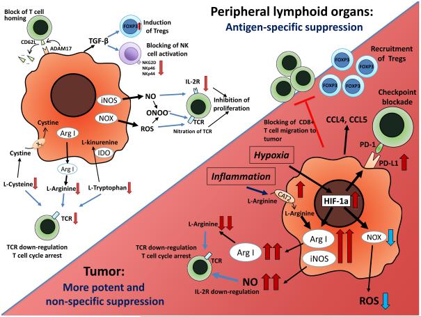
Figure 1. MDSC function in tumor site and peripheral lymphoid organs.
In peripheral lymphoid organs, immune suppression by MDSC is mainly antigen-specific, contact-dependent and utilizes several major pathways, including the production of reactive nitrogen and oxygen species (NO, ROS and PNT), elimination of key nutrition factors for T cells from the microenvironment (L-arginine, L-tryptophan and L-cysteine), disruption of homing of T cells (through the expression of ADAM17), production of immunosuppressive cytokines (TGF-β, IL-10), and induction of T regulatory (Treg) cells. After migration to the tumor, MDSC are exposed to inflammatory and hypoxic tumor microenvironment. This results in significant HIF-1α-mediated elevation of Arg1 and iNOS and downregulation of ROS production, upregulation of inhibitory PD-L1 on MDSC surface, and production of CCL4 and CCL5 chemokines attracting Tregs to the tumor. Overall, these alterations result in more potent non-specific immunosuppressive activity of MDSCs inside the tumor.

