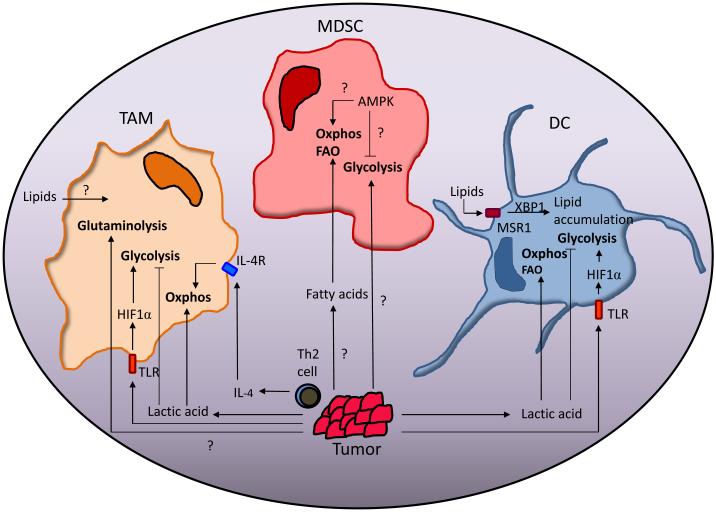
Figure 3. The effect of the tumor microenvironment on the metabolism of myeloid cells.
Lactic acid produced by tumor cells and IL-4 produced by Th2 cells in the TME can drive the metabolism of TAM and TADC towards oxidative phosphorylation (oxphos) while inhibiting glycolysis. Lipids are known to have a role in negative regulation of TADC function. MDSC in peripheral tissue have decreased rates of oxphos and glycolysis compared to tumor-infiltrating MDSC (T-MDSC). Fatty acids derived from the TME drive the metabolism of T-MDSC towards fatty acid oxidation (FAO) and oxphos. Glycolytic rates are also increased in T-MDSC but how the TME influences this process, and the role AMPK plays in this process is unclear.

