Figure 3.
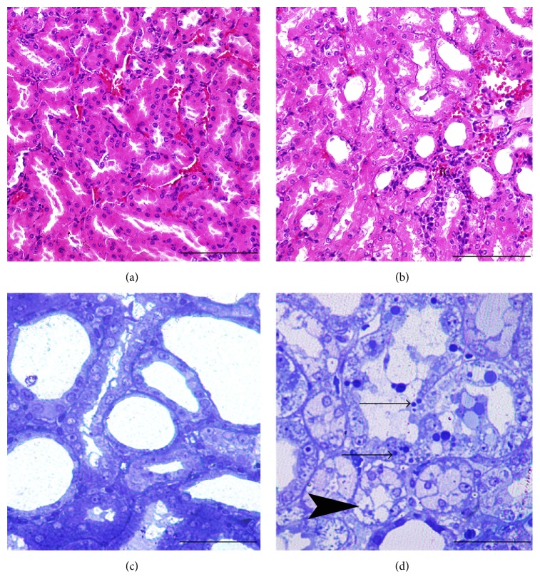
Morphology of renal tubular cells of HRs ((a), (c)) and CIN + DRs ((b), (d)). Histopathological changes such as mononuclear and polymorphonuclear leukocytes infiltration of interstitial cells (IC) of CIN + DRs (Figure 3(b)). Apoptotic tubular cells with membrane-bound apoptotic bodies (black arrow) and quite noticeable glucogenic vacuolization (arrow head) in tubular cells of CIN + DRs (Figure 3(d)). Note: histological micrographs were stained with H&E and had scale bars with 500 μ (Figures 3(a) and 3(b)). Histological micrographs were stained with toluidine blue and had scale bars with 150 μ (Figures 3(c) and 3(d)). HRs: healthy rats; CIN + DRs: diabetic rats with contrast-induced nephropathy.

