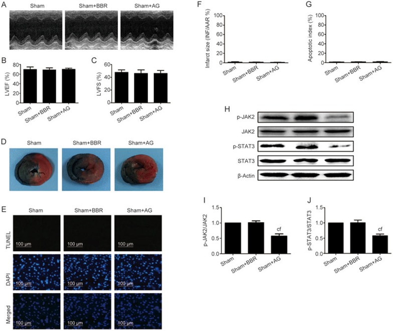
Figure 2.
BBR or AG490 treatment had no significant toxic effect on sham-operated rat hearts. Normal rats pretreated with BBR or AG490 were subjected to the sham operation. After 24 h of reperfusion, echocardiography was performed. After 6 h of reperfusion, the myocardial infarct size and apoptosis index were evaluated. (A) Representative M-mode echocardiographic images. (B) Left ventricular ejection fraction (LVEF). (C) Left ventricular fractional shortening (LVFS). (D) Representative heart section images. The Evans blue-stained areas (blue) indicate the non-ischemic/reperfused area; the TTC-stained areas (red) indicate ischemic but viable tissue; and the Evans blue/TTC-unstained (negative) areas (white) indicate infarcted myocardium. (E) Representative images of apoptotic cardiomyocytes by TUNEL staining. The green fluorescence shows the TUNEL-positive nuclei; the blue fluorescence shows the nuclei of all cardiomyocytes; original magnification, ×400. (F) The myocardial infarct size expressed as the percentage of area-at-risk. (G) Percentage of TUNEL-positive nuclei. (H) Representative blots. (I) p-JAK2/JAK2 ratio. (J) p-STAT3/STAT3 ratio. BBR, berberine; AG, AG490. The results are expressed as the mean±SEM. n=8/group. cP<0.01 vs the sham group. fP<0.01 vs the sham+BBR group.

