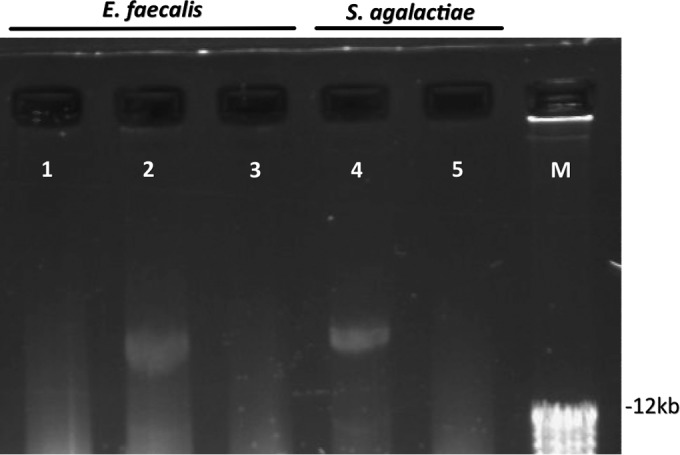
FIG 1.

Plasmid preparations of GBS and E. faecalis strains. Shown are plasmid preparations of E. faecalis and GBS strains separated by agarose gel electrophoresis (0.8% gel). Lanes: 1, E. faecalis strain BSU386 (wild-type strain without HLGR); 2, E. faecalis strain BSU580 (wild-type strain BSU386 after transformation with plasmid preparation from S. agalactiae strain BSU452, displaying HLGR); 3, E. faecalis strain BSU720 (E. faecalis strain BSU580 after plasmid curing and loss of HLGR); 4, S. agalactiae strain BSU452 (patient isolate displaying HLGR); 5, S. agalactiae strain BSU729 (S. agalactiae strain BSU452 after plasmid curing and loss of HLGR). M, molecular size marker.
