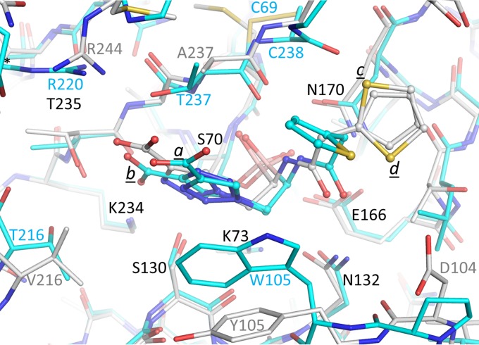
FIG 6.
Superposition of SHV-1- and KPC-2-bound S02030 structures. The protein atoms are shown in stick representation and the ligands are shown in ball-and-stick representation. The KPC-2–S02030 structure is depicted with the carbon atoms in cyan; the SHV-1–S02030 structure is depicted with the carbon atoms in white. The Cα atoms of residues 68 to 84, 121 to 140, 167 to 172, and 233 to 238 of SHV-1 were superimposed on the identical residues of KPC-2, yielding a root mean square deviation (RMSD) of 0.51 Å.

