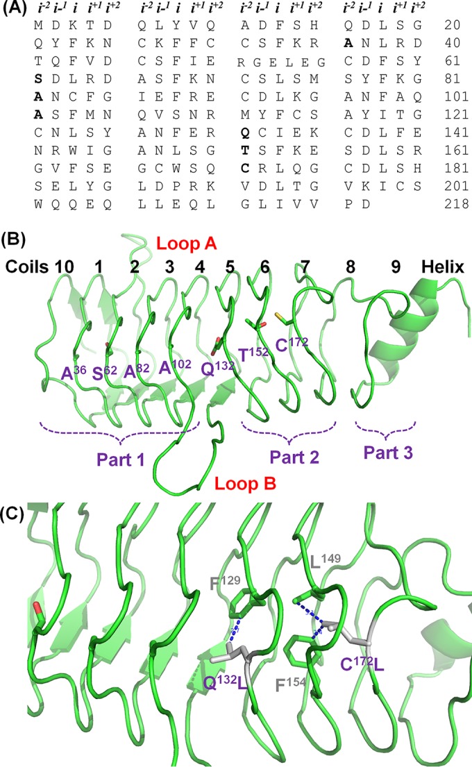
FIG 1.

Amino acid sequence and structural representation of QnrVC7 and specific mutant proteins. (A) Tabular array of QnrV7 amino acids grouped by pentapeptide repeats, with coils along the vertical axis and faces along the horizontal axis; i−2 residues characterized in this study are in bold. (B) Overall structure of QnrVC7 and its organization. The sites at the i−2 position of face 4 are indicated in purple. (C) Amino acid substitutions that led to an increase in protective activity of QnrVC7. The potential hydrophobic interactions that may enhance the stability of QnrVC7 are indicated in gray and purple.
