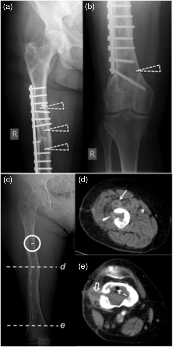
Figure 1.

Chronic femoral osteomyelitis demonstrated by plain radiographs and computed tomography in 2007. Anterior-posterior plain film projections (a and b) show the orthopedic hardware and several radiolucencies within the femur (open dashed arrowheads). Scout image of the computed tomography (c) shows the remnant of a screw (circle); location of axial images performed with intravenous contrast are marked with lines (d and e). Image (d) demonstrates an abscess formation ventral to the midportion of the femur within the vastus intermedius muscle (arrow), extending to the bone marrow (arrowhead) through a borehole defect. (e) In the distal femur, intramedullary gas (dashed arrow) within a fluid collection is shown, extending through a lateral cortical defect and a sinus tract (open arrow) to the skin.
