
Fig. 1.
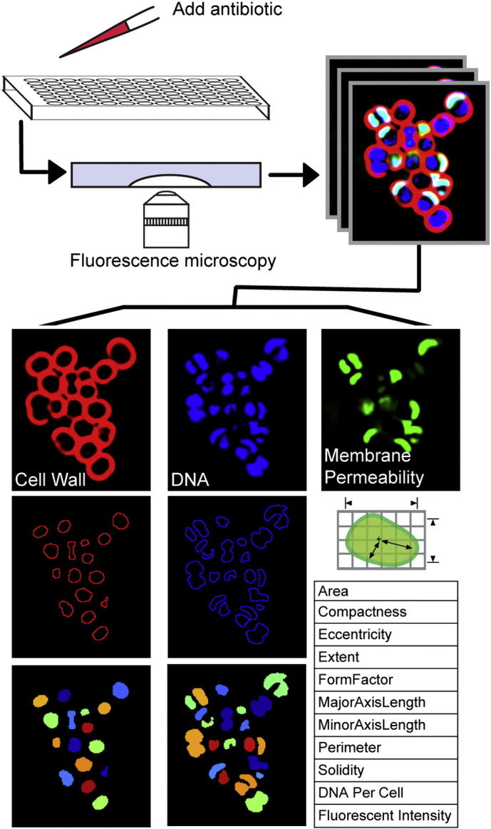
BCP methodology and analysis. Each antibiotic was added to exponentially growing cells and samples were collected for imaging hourly. Changes in cytological parameters were measured using CellProfiler. For each strain, three sets of profiles (each represented by three images), were obtained over three different days.