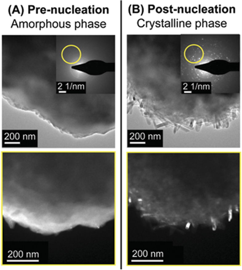
Figure 2. Amorphous to crystalline phase transition.
(A) Prior to nucleation the precursor remains amorphous as can be seen from the featureless diffraction pattern in the inset. The region of the diffraction pattern encircled in yellow originates from the features highlighted in the image below (Selected Area Electron Diffraction, or SAED), providing a visual view of the amorphous character of the precursor. (B) A multitude of bright spots appear in the diffraction pattern after the NWs have been nucleated. The SAED study reveals crystalline NWs are responsible for the selected set of diffraction spots once the amorphous-crystalline phase transition has been completed.

