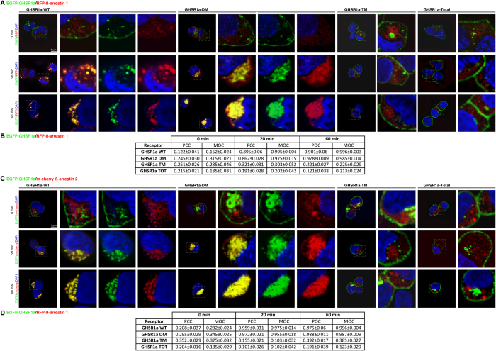
Figure 3. The phospho-acceptor residues in C-terminal tail of GHSR1a-WT govern both binding and trafficking patterns of ß-arrestins.
(A) Trafficking of the RFP-tagged ß-arrestin 1 with the EGFP-tagged GHSR1a-WT or the mutants GHSR1a DM, GHSR1a -TM or GHSR1a TOTAL expressed in HEK 293 cells. (B) Correlation measurements between RFP-tagged ß-arrestin 1 with the EGFP-tagged GHSR1a-WT or the mutants were made on a single cell using two coefficients: the PCC and MOC. (C) Trafficking of the m-cherry-tagged ß-arrestin 2 with the EGFP-tagged GHSR1a-WT or the mutants GHSR1a DM, GHSR1a -TM or GHSR1a Total expressed in HEK 293 cells. (D) PCC and MOC correlation coefficients were evaluated as the measurement of colocalization between m-cherry-tagged ß-arrestin 2 with the EGFP-tagged GHSR1a-WT or the mutants. In (A,C) confocal images show the localization of the ß-arrestin and the GHSR1a before and following stimulation with ghrelin (100 nM) for 20 or 60 minutes. Receptor fluorescence is shown in green and ß-arrestin fluorescence in red. Co-localisation of ß-arrestin and the receptor is shown in yellow when images are merged. Results are representative of three similar experiments. In (B,C) The data are expressed as the mean ± SEM.

