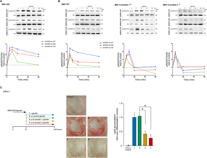
Figure 6. Functional relation between the GHSR1a-associated ß-arrestin-scaffolded complex and the Akt activity.
(A) HEK 293 cells were transiently transfected with GHSR1a-WT or mutants and stimulated with ghrelin (100 nM) for the indicated times. Samples of cell lysates were separated by SDS PAGE and immunoblots were performed using anti pAkt (S473) or total Akt antibodies. The levels of pAkt (S473) were quantified by densitometry, normalized to total Akt, and expressed as the fold change relative to the unstimulated cells. (B) The MEF WT, ß-arrestin 1−/− and ß-arrestin 2−/− cells treated as in (A) and the levels of pAkt (S473) were expressed as the fold change relative to the unstimulated cells. In (A,B) immunoblots are representative of three independent experiments and data are expressed as the mean ± SEM. (C) Effect of siRNA depletion of ß-arrestins on terminal adipogenesis in 3T3-L1 cells. After induction for 3 days under treatment with IBMX (0.5 mM), DEX (25 μM), and ghrelin (861 nM) in DMEM/10% FBS, the cells were transfected with specific ß-arrestin 1 or 2 siRNAs and maintained for 3 days in the presence of ghrelin (172 nM) in DMEM/10% FBS. The cells were stained with Oil red O and the lipid droplet accumulation was analyzed using the spectrophotometric absorbance at 520 nm. The results are expressed as the fold change in lipid accumulation relative to the differentiated controls.

