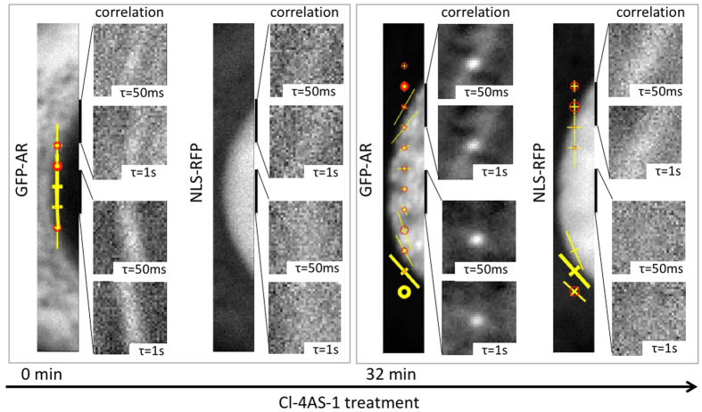
Figure 4. Anisotropic GFP-AR movement results from structural confinement and slow kinetics.
Before adding Cl-4AS-1, there was no significant directionality of GFP-AR along the nuclear envelope. After Cl-4AS-1 treatment, GFP-AR showed strong directionality parallel to the nuclear envelope. In contrast, the simultaneously acquired NLS-RFP signal showed lower level of correlation in both before and after agonist treatment cells due to its fast diffusive nature. Note that the nuclear envelope boundary and the intensity differences are not the sole reasons for the observed confined anisotropic movement, as both NLS-RFP and GFP-AR showed distinct cytoplasmic/nucleus distribution.

