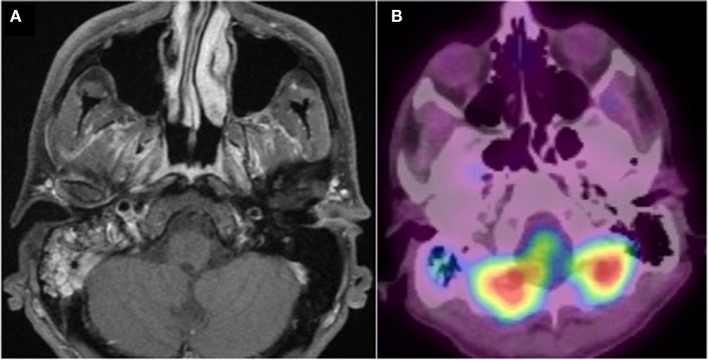
Figure 2.
Post treatment imaging. (A) Gadolinium-enhanced axial T1 weighted fat-suppressed MRI. Minimal persistence of gadolinium enhancement in the right mastoid. (B) PET CT fused image. Good aeration and minimal persistence of 18 FDG uptake in the right mastoid. No other associated bone or tissue anomaly.

