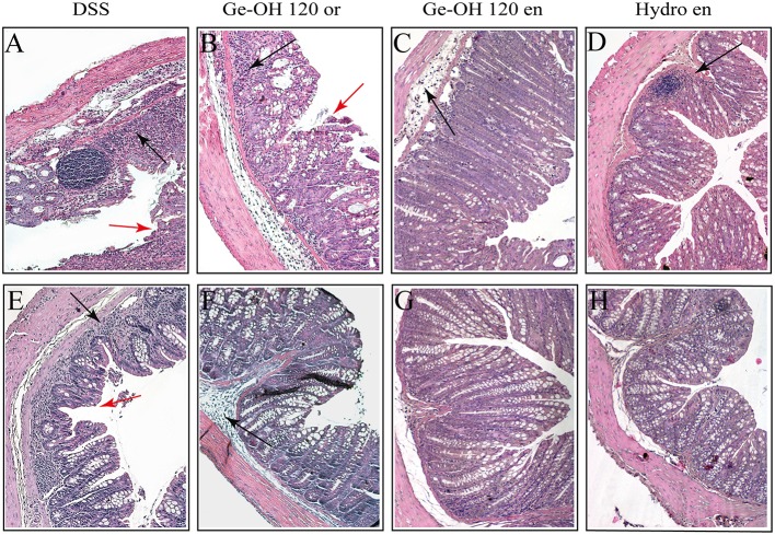
Figure 4.
Differences in histological architecture induced by Ge-OH 120mg kg(−1) and hydrocortisone 2.5 mg kg(−1) during the experimental colitis. Colon specimens were collected from mice on days 25 (A–D) and 37 (E–H). Histopathological changes in individual crypts are shown in representative hematoxylin and eosin-stained sections. Red arrows indicate loss of crypt architecture associated with epithelial damage and flattened villi while black arrows indicate leukocyte infiltration (Magnification: 10X; bar = 100 μm).

