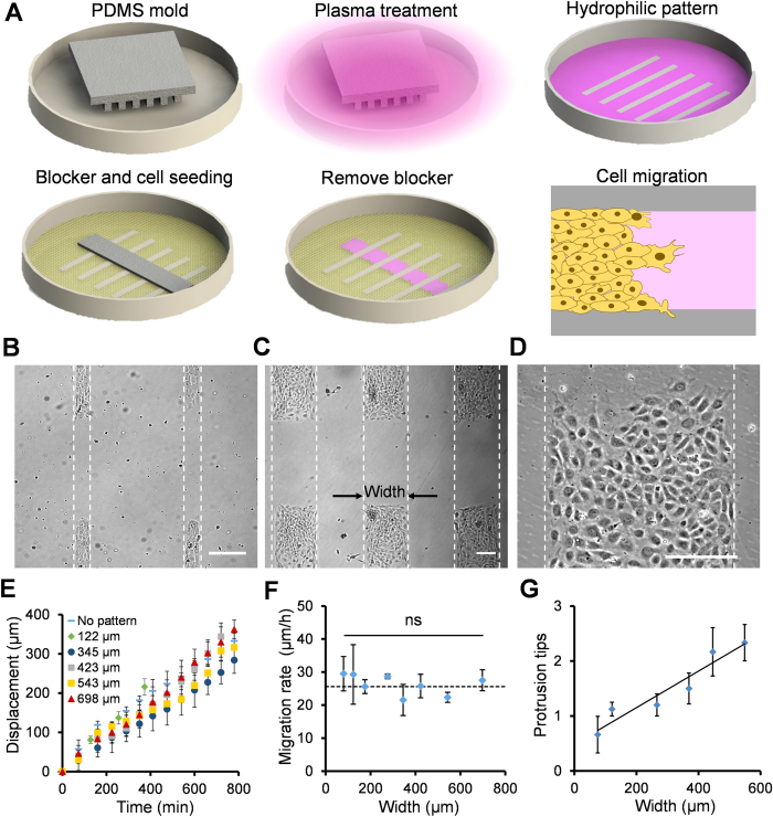
Figure 1. Endothelial collective migration in geometrically confined microenvironments.
(A) Schematic of the plasma lithography modulated wound healing assay. In this assay, hydrophilic cell adhesion patterns are created by physical shielding of plasma treatment with a PDMS mold. A PDMS blocker is placed in the plasma lithography patterned polystyrene dish before cell seeding. After a cell monolayer is formed, the PDMS blocker is removed to create a cell free region for studying collective migration. (B–D) Representative images of rectangular cell patterns with different widths. Scale bars, 200 μm. (E) The displacement of cells in patterns with different widths. (F) The migration rate of cells in patterns with different widths. The dotted line indicates the migration rate without plasma lithography patterning. Data represent mean ± s.e.m. (ns: not significant, ANOVA). (G) The average number of protrusion tips for cells in patterns with different widths. The solid line represents the best fit line. Data represent mean ± s.e.m.

