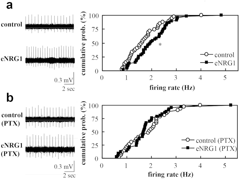
Figure 2. Effects of perinatal eNRG1 administration on firing rates of dopaminergic neurons in midbrain slices.
Mean firing rates of single units are compared in the presence (b) or absence (a) of 50 μM picrotoxin (PTX). Single units were recorded in vitro in the anterior region of medial terminal nucleus of the accessory optic tract. Typical single unit traces are displayed (left panels in a, b). (a) Cumulative probability distributions of firing rates are compared between control and eNRG1 groups in the control condition (n = 39 cells from 4 control mice and n = 42 cells from 5 eNRG1-pretreated mice). (b) Cumulative probability distributions of firing rates in the presence of PTX are compared (n = 33 cells from 4 control mice, n = 46 cells from 5 eNRG1-pretreated mice). *p < 0.05, Mann–Whitney U tests. Note; Coefficient variation of interspike intervals was 6.2 ± 0.6% for control, 4.5 ± 0.5% for eNRG1, 4.5 ± 0.5% for control plus PTX, and 4.7 ± 0.5% for eNRG1 plus PTX (F3,157 = 3.2, p = 0.023, ANOVA, post-hoc; p < 0.05 for a PTX effect in control group).

