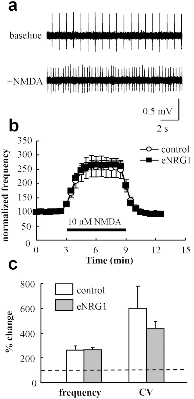
Figure 7. Effects of perinatal eNRG1 pretreatment on NMDA receptor sensitivity in dopaminergic neurons.

Midbrain slices were prepared from control and eNRG1-pretreated mice and single unit activities were monitored from dopaminergic neurons in the presence or absence of 10 μM NMDA. (a) Typical single unit traces are displayed. (b) The time dependency of the NMDA effects is displayed. Mean firing rates before NMDA application were set to 100% in each cell (n = 13 cells from 4 control mice, n = 17 cells from 4 eNRG1 pretreated mice). (c) Mean frequency and coefficient of variation (CV) of interspike intervals before NMDA application were set to 100% (a horizontal broken line) and compared with those after NMDA application (from 4 min to 6 min) (p = 0.47 for frequency; p = 0.71 for coefficient variation, Mann–Whitney U test).
