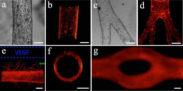
Figure 2.
Endothelial cell-lined lumens and possible morphologies. (a) Image of endothelial cell-lined lumen and (b) 3D volume reconstruction of the biomimetic blood vessel using F-actin staining (phalloidin). (c) Bright field image of bi-branched lumen and (d) multi-branched (tertiary branches) with phalloidin staining. (e) Angiogenesis via vascular endothelial growth factor (VEGF) gradient from source (dashed blue lines). (f) Cross-section of circular lumen lined with endothelial cells (phalloidin staining). (g) Bifurcated lumen lined with endothelial cells. Bar=150μm.

