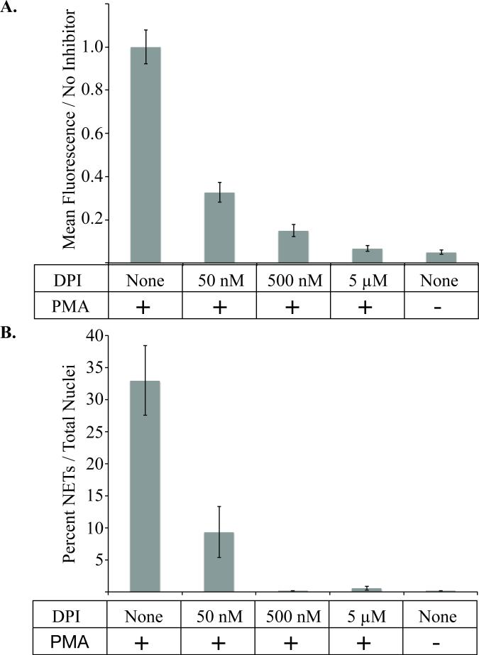
Figure 6. Diphenyleineideom (DPI) inhibits PMA induced neutrophil activation.
A, The mean fluorescence intensity of cells treated with 1μM PMA for 30 minutes with or without the addition of varying concentrations of DPI inhibitor. Fluorescence is measured by dihydrorhodamine-123 (DHR) signal relative to positive control cells activated with 1μM of PMA without inhibitor. There is a statistically significant decrease in formation of reactive oxygen species measured by DHR signal as determined by oneway ANOVA (p-value < 0.001). B, The percent of NETs produced in 3 hours after activation with 1μM PMA in the presence or absence of the indicated concentration of DPI. All data was repeated in duplicate or in triplicate at least three times.

