
Fig. 4.
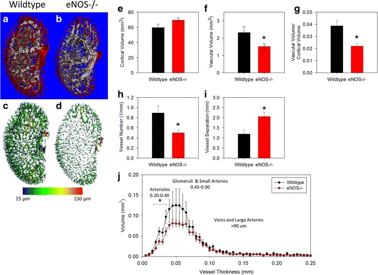
eNOS-/- mice have less perfusable vessels in the renal cortex. Representative images of the renal vasculature in the renal cortex illustrate the perfusion deficiency eNOS-/- mice (b and d) compared to WT mice (a and c). Three-dimensional quantification revealed the deletion of eNOS did not alter (e) the total cortical volume. However, (f) the cortical vascular volume, (g) the cortical vascular volume/cortical volume and (h) the cortical vascular number in eNOS-/- kidneys was significantly lower than in WT kidneys. i Conversely, vessel separation (the mean distance between vessels) was significantly greater in the cortex of eNOS-/- mice. j Histograms illustrating the cortical vascular volume at each possible thickness were created and the total volume of perfused vessels at each given thickness was compared between genotypes. This analysis demonstrated a deficit in the number of perfused vessels in the range of 20–40 μm in thickness in the eNOS-/- renal cortex compared to WT. All data are mean ± SEM. *p < 0.05 vs. Wildtype by Student’s t-test