Fig. 2.
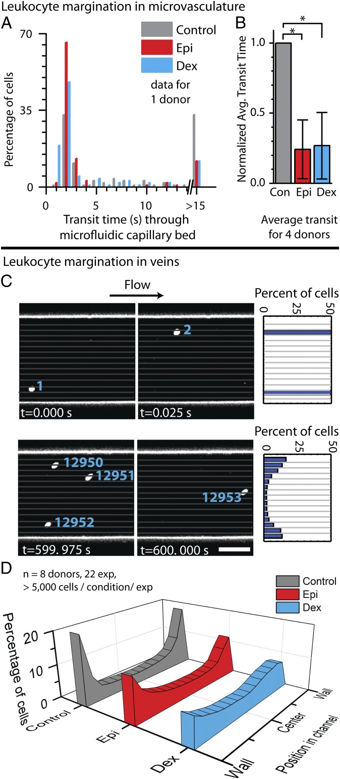
In vitro exposure to glucocorticoids or catecholamines alter granulocyte trafficking in microfluidic systems. (A) Compared with untreated controls, isolated leukocytes from one representative healthy donor treated with the glucocorticoid dexamethasone (Dex) or the catecholamine epinephrine (Epi) have shorter transit times with less propensity to cause microchannel obstruction within the microfluidic capillary bed model (P < 1 × 10⁻⁵ for each condition via Mann−Whitney test). (B) Aggregate results from four donors further show that leukocytes exposed to dexamethasone or epinephrine have shorter transit times than control leukocytes (P < 1 × 10⁻⁶ via Mann−Whitney test). (C) Leukocyte demargination within the microfluidic venous model is tracked by imaging stained leukocytes within whole blood in microfluidics via confocal videomicroscopy. The leukocyte position with respect to the microfluidic wall was calculated using a custom MATLAB imaging processing script. (D) The histogram shows leukocyte locations (perpendicular to flow) pooled from eight donors over 22 experiments, with >5,000 cells analyzed per condition per experiment at physiological venous wall shear stress of 1 dyne/cm2. Compared with untreated controls, leukocytes treated with dexamethasone strongly demarginated, and leukocytes treated with epinephrine showed a similar but less pronounced demargination.

