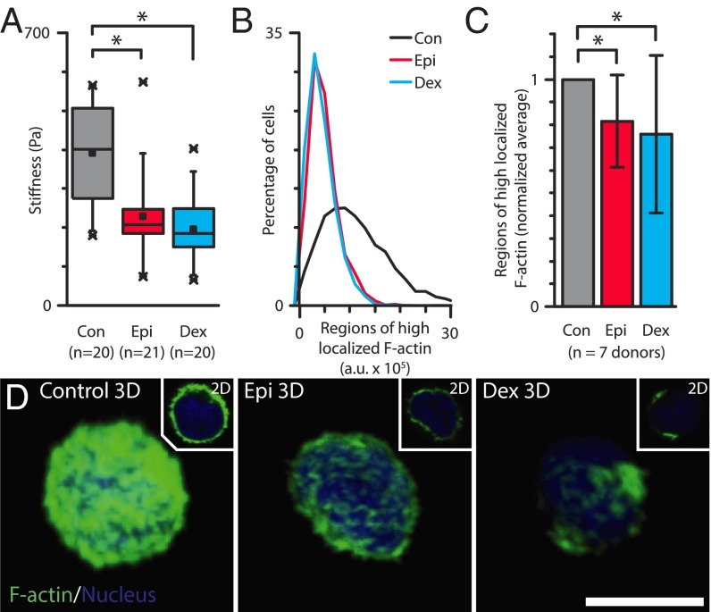
Fig. 3.
Granulocytes exposed to glucocorticoids and catecholamines are mechanically softer due to reorganization of the cortical actin cytoskeleton. (A) AFM measurements of granulocytes from a single donor exposed to physiologically relevant doses of glucocorticoids (Dex) and catecholamine (Epi) reveal a significant reduction in cellular mechanical stiffness compared with untreated controls (P < 0.0001 and 0.001, respectively.) Box plot denotes 25th and 75th percentile; “x” denotes first and 99th percentile, with central dot representing the mean value. (B) Flow cytometry analysis of phalloidin staining reveals significant reductions in localized, polymerized actin in granulocytes exposed to dexamethasone and epinephrine for one representative donor. (C) Aggregate flow cytometry results from six donors further show reduced localized actin in treated leukocytes compared with untreated leukocytes (P < 0.01 via Mann−Whitney test.) (D) Confocal microscopy images show less polymerized actin in granulocytes after exposure to epinephrine and dexamethasone. (Scale bar, 5 μm.)

