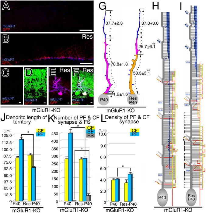
Fig. 5.
Restored refinement in CF and PF innervation after lentiviral transfection of mGluR1α in mGluR1-KO PCs. (A–F) Triple fluorescent labeling for GFP (red), mGluR1α (blue), and calbindin (green) in mGluR1α/GFP-transfected mGluR1-KO cerebellum at P40. Untransfected (A, C, and D) and transfected (B, E, and F) cerebellar portions are shown. (G and H) Reconstructed dendritic innervation in mGluR1-KO (G) and rescued (H) PCs. One of the three PCs examined in mGluR1-KO and mGluR1α-rescued PCs is illustrated here, with the rest being shown in Fig. S7. (I) The mean path lengths of the three dendritic domains (µm, mean ± SD). (J–L) Bar graphs showing the mean path lengths of CF and PF territories (J), the mean number of CF synapses, PF synapses, and free spines (K), and the mean density of CF and PF synapses (L). Note the emergence of free spines in the CF territory of rescued PCs (black squares in H and gray column in K). See legends for Figs. 2 and 3. (Scale bars, 100 µm in A and B; 10 µm in C–F.)

