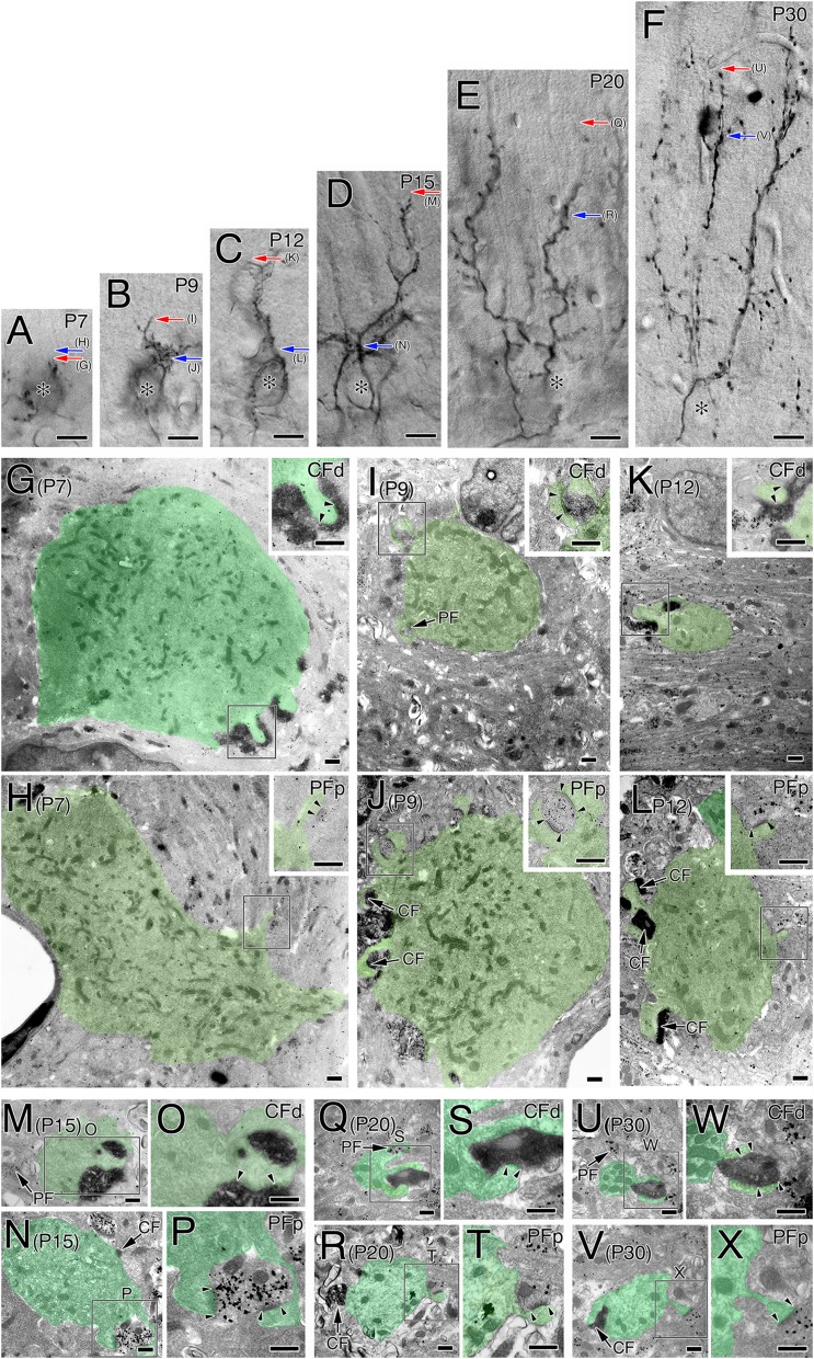
Fig. S1.
(A–F) Bright-field micrographs of BDA-labeled CFs used for ultrastructural analysis from P7 to P30. For ultrastructural reconstruction, we selected BDA-labeled CFs whose trajectories could be traced from PC somata to the tips of CFs. Red and blue arrows indicate approximate portions of the most distal CF synapses (CFd) and the most proximal parallel fiber synapses (PFp) shown in G–X. After taking these micrographs, sections were subjected to pre-embedding silver-enhanced immunogold microscopy for VGluT1 to label PF terminals, and embedded in Epon812. Asterisks indicate PC somata. (G–X) Electron micrographs showing the most distal CF synapses and the most proximal PF synapses at P7 (G and H), P9 (I and J), P12 (K and L), P15 (M–P), P20 (Q–T), and P30 (U–X). Boxed regions in G–L are enlarged in Insets, whereas those in M, N, Q, R, U, and V are enlarged in O, P, S, T, W, and X, respectively. Insets in G, I, and K and closer views of O, S, and W show the most distal CF synapses (CFd) labeled with BDA tracer (diffuse DAB precipitates). Insets in H, J, and L and closer views of P, T, and X show the most proximal PF synapses (PFp) labeled for VGluT1 (metal particles). Arrows indicate the coexistence of CFd with PF synapses or of PFp with CF synapses (CF). Pairs of arrowheads indicate both edges of the postsynaptic density. PC dendrites or somata are pseudocolored in green. Note reduced calibers of CFd- and PFp-associated PC dendrites because of distal elongation of CF territory or distal retraction of PF territory, respectively, during early postnatal period. Also note that the most distal CF synapses at P7 are observed at apical portions of PC somata or somatodendritic border. (Scale bars, 10 μm in A–F; 500 nm in G–X.)

