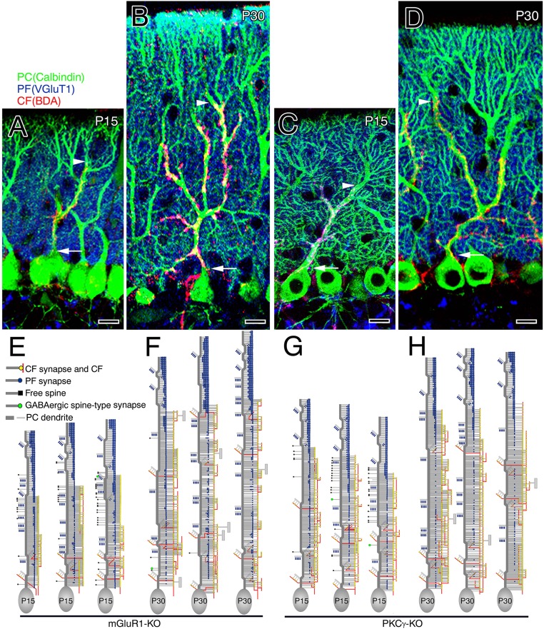
Fig. S6.
CF and PF innervation in mGluR1-KO (Left) and PKCγ-KO (Right) mice. (A–D) Triple fluorescent labeling for CFs (red, BDA tracer labeling), PF terminals (blue, VGluT1 immunofluorescence), and PCs (green, calbindin immunofluorescence) at P15 (A and C) and P30 (B and D) in mGluR1-KO (A and B) and PKCγ-KO (C and D) mice. Arrowheads indicate the tip of the CF projection, whereas arrows indicate the somatodendritic border of PCs. (Scale bars, 10 μm.) (E–H) Reconstructed dendritic innervation in three PCs at P15 (E and G) and P30 (F and H) in mGluR1-KO (E and F) and PKCγ-KO (G and H) mice. See legends for Figs. 1 A–F and 2 A–F.

