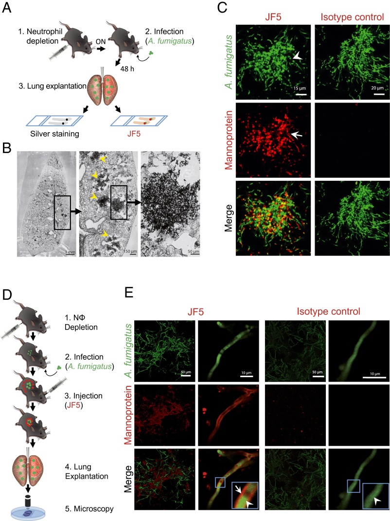
Fig. 1.
Binding specificity of JF5 to A. fumigatus in infected mouse lungs. (A) Experimental workflow for the in situ binding of the mAb JF5: (1) Neutrophil depletion by i.p. injection of 100 µg anti-Gr-1 antibody (clone RB6-8C5) 17 h before infection. (2) Intratracheal infection with A. fumigatustdTomato. (3) After 48 h, lung explantation, fixation, and slicing followed by in situ staining with either JF5-DyLight650 or methenamine silver. (B) A. fumigatus-infected lung lobe section stained with methenamine silver. Fungal biomass can be detected by its black appearance (yellow arrowheads). (C) Tissue sections of A. fumigatustdTomato (green) infected lungs stained with JF5-DyLight650 (displayed in false color red) or mouse IgG3 isotype control (red). White arrowheads indicate expression of tdTomato in the cytoplasm and JF5-antigen in the hyphal cell wall. (D) Experimental workflow for JF5 binding in vivo: (1) Neutrophil depletion by i.p. injection of 100 µg of anti-Gr-1 antibody (clone RB6-8C5) 17 h before infection. (2) Intratracheal infection with A. fumigatustdTomato. (3) i.v. injection of JF5-DyLight650 or fluorochrome-labeled mouse IgG3 isotype control 24 h after fungal infection. (4) Another 24 h later, lung explantation, followed by (5) in situ confocal microscopy of longitudinally sectioned lung. (E) Micrographs of A. fumigatustdTomato (green) infected lungs treated with JF5-DyLight650 (red) or mouse IgG3 isotype control (red). Blue boxes indicate regions of interest, which are shown at higher magnification in the insets. White arrowheads indicate expression of tdTomato in the cytoplasm and white arrows indicate expression of JF5-antigen in the hyphal cell wall, respectively.

