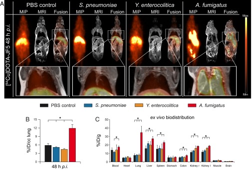
Fig. 2.
[64Cu]DOTA-JF5 as disease-specific PET-tracer for A. fumigatus lung infection. (A) Sagittal maximum intensity projections (MIP), MRI, and fused PET/MRI images of PBS-treated mice and S. pneumoniae-, Y. enterocolitica-, and A. fumigatus-infected mice injected with [64Cu]DOTA-JF5 (48 h after infection). Tracer injection demonstrates highly specific accumulation in A. fumigatus-infected lung tissue compared with bacterial-infected or sham-treated animals. (A, Lower) Lungs of the respective animals. (B) Quantification of the in vivo PET images showed a significantly higher uptake of [64Cu]DOTA-JF5 in the lungs of A. fumigatus-infected animals compared with the lungs of the control groups. (C) The ex vivo biodistribution confirmed the in vivo PET results with significantly higher uptake of [64Cu]DOTA-JF5 in the lungs of A. fumigatus-infected animals compared with the lungs of control animals 48 hpi. The systemic infection at the late stage of the disease in neutropenic and A. fumigatus-infected animals is reflected by higher tracer accumulation in lymphatic and other organs. Black bars, PBS-treated controls (n = 8); blue bars, S. pneumoniae-infected mice (n = 5); orange bars, Y. enterocolitica-infected mice (n = 5); red bars, A. fumigatus-infected mice (n = 5). Data are expressed as the average ± SD (%ID/cc: PET; %ID/g: ex vivo biodistribution), one-way ANOVA, post hoc Tukey–Kramer corrected for multiple comparisons, *P < 0.05.

