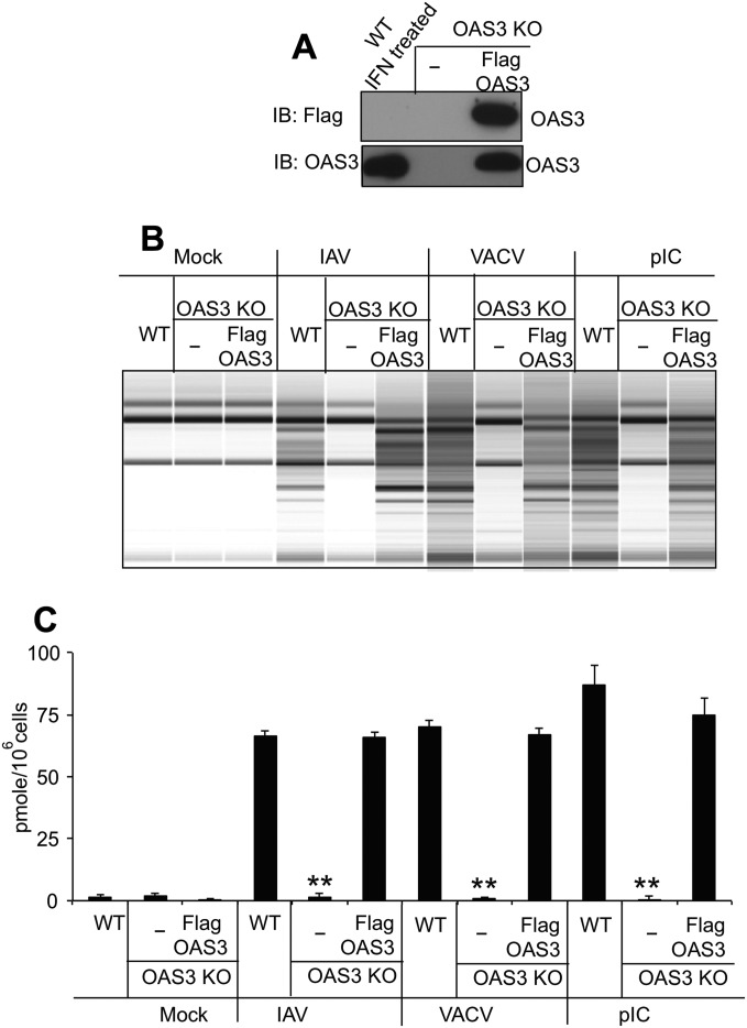
Fig. S1.
Expression of OAS3 protein in OAS3-KO cells restores 2-5A production and RNase L activation. (A) OAS3-KO cells were transfected with p3XFlag-tagged OAS3 cDNA, and a cell line was generated by neomycin selection. Cell lysates were analyzed by electrophoresis followed by immunoblotting with anti-Flag antibody (Upper) and monoclonal antibody against human OAS3 (Lower). (B) OAS3-KO cells and OAS3-KO cells in which OAS3 expression was restored (as indicated) were transfected with pIC (1 μg/mL) or were mock-infected or were infected with IAVΔNS1 (MOI = 10) or VACVΔE3L (MOI = 10). Cells were lysed at 4 (pIC) or 24 (virus) hpi, and total RNA was isolated and monitored for integrity on a Bioanalyzer. (C) OAS3-KO cells and OAS3-KO cells in which OAS3 expression was restored (as indicated) were transfected with pIC (1 μg/mL) or were mock-infected or infected with IAVΔNS1 (MOI = 1) or VACVΔE3L (MOI = 1). Cells were lysed at 4 (pIC) or 24 (virus) hpi, and intracellular levels of 2-5A were determined by FRET assay. **P < 0.0001.

