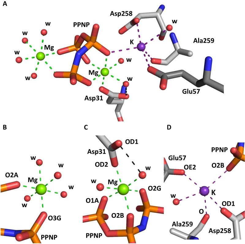
Fig. S2.
Magnesium and potassium ion binding in ligand-bound MATα2. (A) Active site of MATα2 (SAMe+ADO+MET+PPNP) showing two magnesium ions (green) and one potassium ion (purple). (B) Closer view of the first magnesium ion, hexacoordinated by four water molecules and two oxygen atoms, O3G and O2A, from PPNP. (C) Closer view of the second magnesium ion, hexacoordinated by two water molecules and four oxygen atoms, OD2 Asp31, O1A, O2B, and O2G, from PPNP. (D) Potassium ion pentacoordinated by water and four oxygen atoms originating from PPNP (O2B) and residues Asp258 (OD1), Ala259 (O), and Glu57 (OE2). PPNP and MATα2 residues are shown as sticks color-coded according to atom type (gray, C; red, O); water molecules are shown as red spheres. Coordination bonds for magnesium ions are indicated by green dotted lines; those for potassium, by purple dotted lines. Black dotted lines represent hydrogen bonds.

