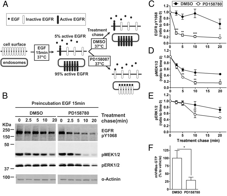
Fig. 4.
EGFR kinase activity is required for sustained MEK and ERK activation. (A) Schematics of the experimental protocol used in B–F. (B) HeLa/mV-HRas cells were incubated with 4 ng/mL EGF at 37 °C for 15 min. DMSO (vehicle) or PD158780 (50 nM) was then added to cells, and the cells were further incubated at 37 °C for indicated periods of time in the same medium (treatment chase). Cell lysates were probed by Western blotting with antibodies to EGFR pY1068, pMEK1/2, pERK1/2, and α-actinin (loading control). (C–E) Graphs show mean values of band intensities normalized to the amount of α-actinin (±SEM; n = 8) exemplified in B and presented as percentage of the mean value at time “0” (after the 15-min incubation with EGF but before the chase treatment). (F) HeLa/mV-HRas cells were incubated with EGF for 15 min, and then with DMSO or PD158780 for additional 5 min as described in B. The amount of mV-HRas⋅GTP was measured using the GST-RBD pull-down assay as in Fig. 1. Bar graphs show mean band intensities (±SEM; n = 5). Paired t tests were performed. *P < 0.05, **P < 0.01, and ***P < 0.001.

