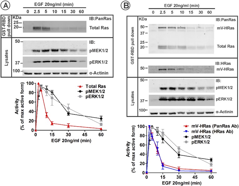
Fig. S1.
Kinetics of Ras and MEK/ERK activity in parental HeLa and HeLa/mVenus-HRas HeLa cells stimulated with 20 ng/mL EGF. (A) Serum-starved HeLa cells were incubated with 20 ng/mL EGF at 37 °C for indicated times. GTP-loaded Ras was pulled down from cell lysates with GST-RBD. GST pulldown and aliquots of the lysates were electrophoresed, and probed by Western blotting for Ras (pan-Ras antibody) in pulldowns, and phosphorylated MEK1/2 (pMEK) and ERK1/2 (pERK), and α-actinin (loading control) in lysates. Western blot images of a representative experiment are shown. Graphs represent mean band intensities normalized to the amount of α-actinin (±SEM; n = 4) expressed as percentage of the maximum high-signal intensity for each signaling protein during the time course. (B) Serum-starved HeLa/mV-HRas cells were stimulated with 20 ng/mL, and the activities of mV-HRas, MEK1/2, and ERK1/2 were measured as described in A. GST pulldown was also probed with the HRas antibody. Western blot images of a representative experiment are shown. Graphs represent mean band intensities normalized to the amount of α-actinin (±SEM; n = 4) expressed as percentage of the maximum high-signal intensity for each protein during the time course.

