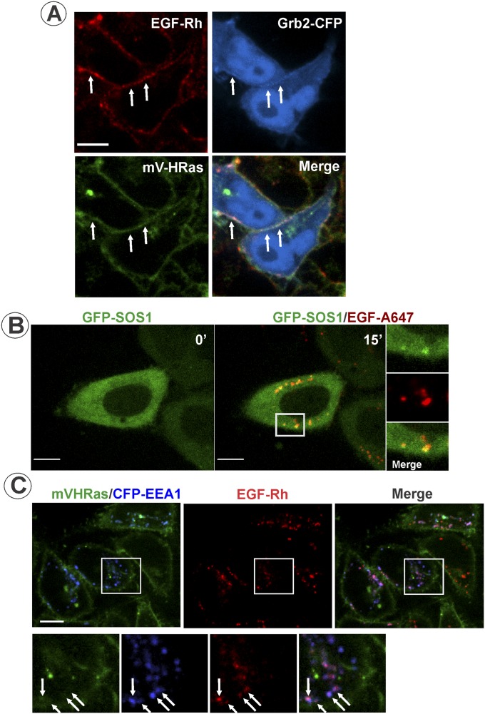
Fig. S3.
Localization of Grb2-CFP, GFP-SOS, and mV-HRas in EGF-stimulated cells. (A) HeLa/mV-HRas cells transiently expressing Grb2-CFP were incubated with 4 ng/mL EGF-Rh for 1 min at 37 °C, and then imaged after 5-min incubation at room temperature to slow down endocytosis. Live-cell 3D images were acquired through 445-nm (CFP; blue), 515-nm (mVenus; green), and 561-nm (rhodamine; red) channels. Arrows indicate examples of the overlap of EGF-Rh, Grb2-CFP, and mV-HRas fluorescence in the plasma membrane (cell edge). (Scale bar: 10 μm.) (B) HeLa cells transiently expressing GFP-SOS1 were incubated with 10 ng/mL EGF-A647. Images were acquired from living cells at 37 °C. Insets represent high-magnification images of the region indicated by the white rectangle. (Scale bar: 10 μm.) (C) HeLa/mV-HRas cells transiently expressing CFP-EEA.1 were incubated with 4 ng/mL EGF-Rh for 15 min at 37 °C. Three-dimensional images were acquired through 445-nm (CFP; blue), 515-nm (mVenus; green), and 561-nm (rhodamine; red) channels from living cells at room temperature to reduce endosome mobility. Insets represent high-magnification images of the region indicated by the white rectangle. Arrows indicate examples of EGF-Rh and CFP-EEA.1 colocalization in endosomes. (Scale bar: 10 μm.)

